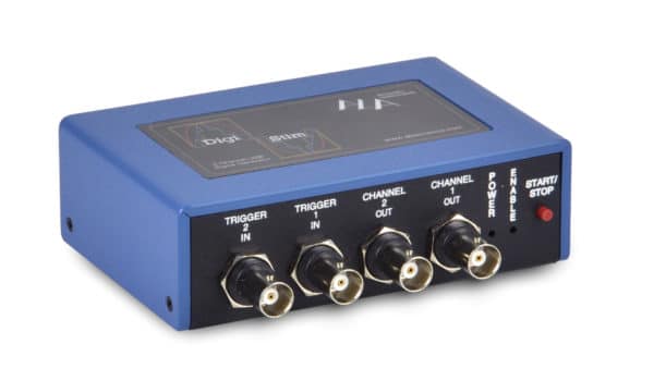
The ELECTROPORATOR is made for electroporation of single cells in slices or cell cultures, e.g. for transfection of DNA. The single cell electroporator is an analog device that converts any input signal into a proportional output signal. The ELECTROPORATOR comes with a built-in audio monitor, a resistance measuring system and a footswitch. For generating the necessary electroporation waveform, we offer the combination with the DigiStim, which is a small USB-programmable signal generator. Read more…
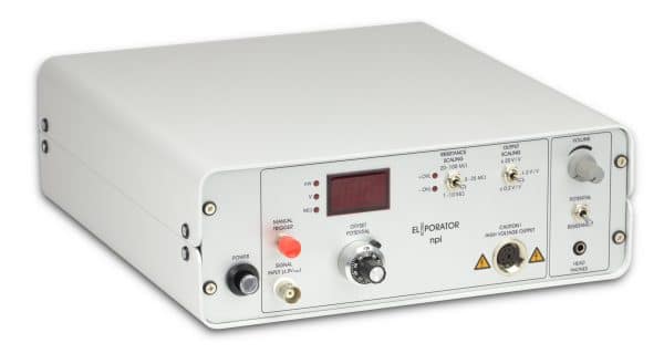
Yes, the ELECTROPORATOR works well with all major data acquisition hardware and software (Spike2, Signal, pClamp, Patchmaster, WinEDR, WinWCP). It can be bundled with the DigiStim into a stand-alone system.
The pipette tip is moved very close to the cell’s membrane. To bring the pipette directly in front of the cell membrane, the user uses the audio signal dependent on the resistance at the pipette tip. At the membrane, the value and thus the sound pitch increases rapidly. Current or voltage pulses are applied to the membrane, which causes the membrane to form small holes in the vicinity of the pipette tip. Charged molecules (DNA, dyes, etc.) can then move along the electrical field into the cell. In contrast to electroporation in a cuvette, this also works with lower voltages: the distance is also lower, resulting in a similar electric field.
Juxtacelular filling or juxtacellular labelling is a technique that allows labelling of extracellularly recording neurons. It is especially useful for structure-function correlation studies in living tissue and intact neural networks. The working principle is to iontophorese tracer molecules into the neuron under constant electrophysiological monitoring. (see doi:10.1007/978-1-60327-202-5_3)
To cover a wide range of electrode resistances and the large difference in resistance between bath, tissue and cell proximity, the resistance measuring circuit has several ranges, if the measuring range is exceeded or undercut, the unit indicates this with “+/-OVER” LEDs and the user switches one range further. This results in a ~100kΩ – ~1MΩ precision, depending on the range.